
Files
Download Full Text (920 KB)
Description
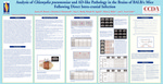
Alzheimer’s disease (AD) is an age-related progressive neurodegenerative disorder and the most common form of dementia. The pathology in the central nervous system (CNS) impairs memory and cognition, hindering the capabilities and the quality of life of the individual. This project continues studying the role of infection and Alzheimer’s disease, as previous studies in this laboratory have done, and contributes to the overall understanding of the possible causes of this disease. In this study, BALB/c mice were infected, via direct intracranial injection, with a respiratory isolate (AR-39) of Chlamydia pneumoniae. Their brains were analyzed at 7 and 14 days post-infection, via immunohistochemistry, for the presence of C. pneumoniae, amyloid deposits and activated glial cells. The goal of this project was to measure the location and degree of C. pneumoniae burden, amyloid deposition and glial cell activation in the CNS following direct intracranial injection and to compare this data with results obtained from previous studies in this laboratory. We hypothesized that C. pneumoniae antigen and activated inflammatory cells will be observed in the infected mouse brains following direct intracranial injection and Aβ deposition will be observed in areas where inflammation occurs. C. pneumoniae, amyloid deposits and activated glial cells were detected in the brains following direct intracranial infection with C. pneumoniae. In infected mice there was an approximate 3.5-fold increase of C. pneumoniae antigen burden compared to uninfected mice at day 7 and there was an approximate 5.5-fold increase of C. pneumoniae antigen burden compared to uninfected mice at day 14. The burden of C. pneumoniae antigen, in the infected mice, increased 1.009-fold (no change) from day 7 to day 14 post-infection. The amyloid burden in infected mice increased approximately 3-fold compared to uninfected mice at day 7 and increased greater than10-fold compared to uninfected mice at day 14. The burden of amyloid, in the infected mice, increased 7-fold from day 7 to 14. From 7 to 14 days post-infection the C. pneumoniae and amyloid deposits located near the injection site spread distally from this location to other regions of the brain. Global activation of glia was observed in the CNS of infected mice at both 7 and 14 days post-infection. This data confirms that C. pneumoniae is capable of establishing an infection in the CNS. Although deposits were observed, the lack of a substantial amount of amyloid deposits suggested that the generation of deposits may require longer than 14 days following C. pneumoniae infection. As early as 7 days post-infection, inflammation is observed in response to the presence of C. pneumoniae and/or soluble amyloid in the CNS and the contribution of both infection with C. pneumoniae and the presence of soluble amyloid elicit the inflammatory response that presumably precedes and contributes to amyloid deposition
Publication Date
2011
Keywords
Chlamydia pneumoniae, AD-like Pathology, Intra-cranial Infection
Disciplines
Bacterial Infections and Mycoses | Nervous System Diseases | Neurology
Recommended Citation
Barton, Jessica Rachel; Hammond, Christine J.; Brady, Amy L.; Appelt, Denah M.; Balin, Brian J.; and Little, Christopher Scott, "Analysis of Chlamydia pneumoniae and AD-like Pathology in the Brains of BALB/c Mice Following Direct Intra-cranial Infection" (2011). Scholarly Posters. 9.
https://digitalcommons.pcom.edu/posters/9
Included in
Bacterial Infections and Mycoses Commons, Nervous System Diseases Commons, Neurology Commons